
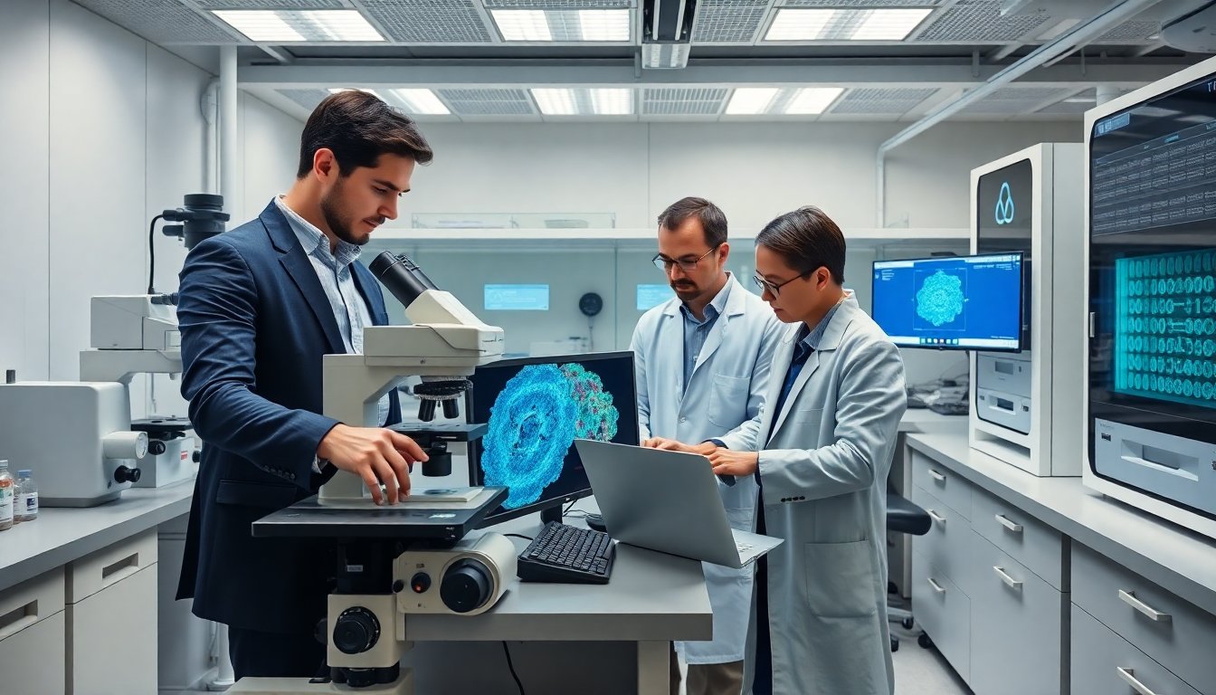

technology revolutionized cell theory development lotsofpower by putting tiny structures into view. Microscopes gave scientists clear images. New detectors and computation gave precise measures. This article shows how optical tools, electron imaging, and computational methods changed cell theory. It highlights key inventions, the shift to live imaging, and the role of data in proving cellular principles.
Table of Contents
ToggleKey Takeaways
- Technology revolutionized cell theory development by enabling detailed visualization of cells through advanced microscopes and imaging techniques.
- Optical and electron microscopy breakthroughs allowed scientists to observe cellular structures, shifting cell theory from hypothesis to proven science.
- Live-cell and super-resolution imaging provided dynamic views of cellular processes, enhancing understanding of cell function and structure.
- Computational imaging and AI automate analysis, increasing precision and reproducibility in studying cells at large scales.
- Big data and quantitative evidence transformed cell theory into a measurable science with rigorous standards and predictive models.
- Advances in hardware, software, and data management collectively empowered researchers to establish robust, testable cellular principles.
Early Microscopy: How Optical Advances Turned Cells From Hypothesis To Visible Units
Early microscopes gave people their first clear view of cells. Antonie van Leeuwenhoek used simple lenses and saw single cells. Robert Hooke used compound lenses and named “cells” after small rooms. Scientists used light, glass, and better stage controls to increase magnification and contrast. Improved illumination let observers see thin tissues and cell walls. Staining methods gave cells distinct colors and boundaries. These methods proved that organisms contained repeating units. The evidence shifted cell ideas from speculation to tested claims. Instruments grew more reliable. Microscope makers improved lens shapes and reduced aberrations. Better mechanical stages allowed careful focusing and repeatable views. These changes let researchers compare samples and replicate results. Reproducible observations supported general statements about cells. Cell theory then stated that organisms consist of cells and that cells form from cells. Optical advances produced the images that made those claims acceptable. Teachers and labs could show cells directly. That visual proof made cell theory part of standard biology. Over decades, optics kept improving. Phase contrast and differential interference provided contrast without stains. Achromatic and apochromatic lenses reduced color errors. These steps increased clarity and confidence in cellular observations.
From Electron Microscopy To Live-Cell And Super-Resolution Imaging: Revealing Subcellular Detail
Electron microscopes revealed structures below the limits of light. Scientists used electron beams to see membranes, ribosomes, and virus particles. These images changed ideas about internal cell structure. Cryo-electron methods preserved native forms. That preservation gave more accurate models of macromolecular arrangements. Later, fluorescent markers let scientists watch proteins inside live cells. Fluorescence methods gave both location and dynamics. Researchers used confocal systems to reduce out-of-focus blur. The arrival of super-resolution tools broke the diffraction limit. Methods such as STED, PALM, and STORM let scientists map structures at tens of nanometers. These maps updated models of organelle architecture and protein complexes. Live-cell super-resolution combined spatial detail with time resolution. This combo changed how scientists test cell function. Researchers could observe signaling, transport, and division at scales that matter. The result was a more detailed and testable cell theory. Imaging technologies also influenced other fields. Sports and tracking systems used microelectronics and sensors for player data. The NHL once removed microchipped gear after early tests, which shows how engineering and validation interact across fields: that removal followed reports about tracking pucks pulled. Engineers applied lessons from instrumentation to biological imaging. Hardware miniaturization and sensor design improved detectors in microscopes. Faster cameras and more stable stages reduced motion blur in live samples. Software improvements handled large image volumes. Together, these tools drove finer, faster, and more reliable cellular observations.
Computational Imaging, AI, And Big Data: Rethinking Cell Theory With Quantitative Evidence
Computational imaging extracts more signal from raw data. Algorithms merge frames, correct drift, and increase resolution. AI models segment images and count organelles automatically. These tools let teams analyze thousands of cells per day. Big data approaches let scientists test population-level claims about cell behavior. Quantitative measures now support statements about growth rates, shape distributions, and protein localization. That evidence tightened links between observation and theory. Researchers used modeling to predict how cell components interact. Models then guided experiments and imaging choices. AI also helped infer unseen structures from partial data. That inference reduced the need for invasive labels. Automated pipelines increased reproducibility. Teams shared code and image sets to let others validate results. Standardized metrics gave clear benchmarks for image quality and analysis performance. These practices made claims about cells more rigorous. Data management tools handled terabytes of image data and kept metadata intact. Cloud computing let teams run heavy analyses without local clusters. Combined, computation and data scale turned qualitative observations into statistical evidence. As a result, cell theory gained more precise statements about variation, mechanism, and scale. Scientists could now say how often a process occurs and under which conditions. That shift moved cell theory from descriptive rules to measurable laws.